100% Native American Owned
Sales, service & parts for healthcare imaging equipment. Multi-vendor certified engineers with decades of experience.
What We Do
New and pre-owned medical imaging equipment from leading vendors. We match your facility with the right technology and budget.
Expert maintenance, repair, and preventive service for your imaging equipment. Multi-vendor trained engineers you can rely on.
Sourcing and supplying parts for a wide range of imaging platforms. Fast turnaround to keep your equipment running.

Welcome to Rose Rock Biomedical
We specialize in providing sales and service for medical imaging equipment, with a well-trained staff of engineers and product specialists carrying many years of experience in healthcare imaging.
We can service and source parts for many different vendor platforms and have been trained across various systems — giving your facility one reliable point of contact.
Get in Touch100% Native American owned and operated — proudly serving the healthcare industry with integrity and community values.
Trained across a wide range of imaging platforms and vendor systems — one team for all your equipment needs.
Our team brings extensive hands-on experience in healthcare imaging — delivering reliable results with minimal downtime.
The people behind Rose Rock Biomedical.

President
Phone 918-617-7795
Fax 479-208-4022
Email evanjones@roserockbiomedical.com

Vice President
Phone 918-575-8895
Fax 479-208-4022
Email tylerfletcher@roserockbiomedical.com
Ready to Get Started?
Our team is ready to help you find the right imaging equipment for your facility.
Contact Us ›Get in Touch
Sales inquiries, service requests, and parts support. We are here to help.
President
Phone 918-617-7795
Fax 479-208-4022
Email evanjones@roserockbiomedical.com
Vice President
Phone 918-575-8895
Fax 479-208-4022
Email tylerfletcher@roserockbiomedical.com
Rose Rock Biomedical is a 100% Native American owned medical imaging company serving healthcare facilities across the region.
CT Scanners
Sales, service & parts for Canon Medical Aquilion CT scanners. Select a model below.

80-row entry-level CT with AI workflow automation and AiCE deep learning reconstruction. Simply delivers.
View Details →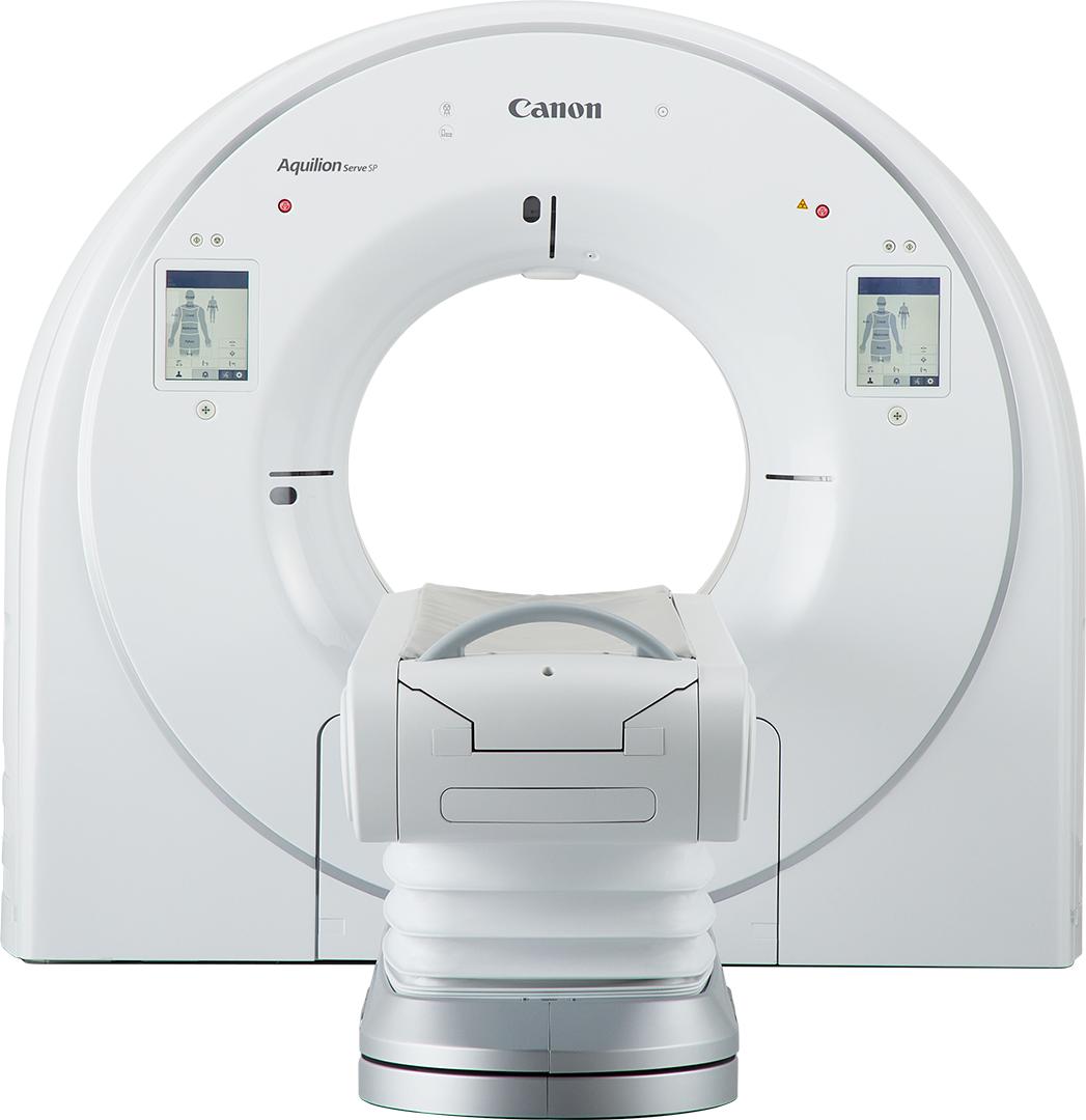
80-row high-performance CT with 72kW generator, 0.35s rotation, and advanced INSTINX AI automation.
View Details →
320-row volume CT with 16cm coverage, spectral CT capability, and PIQE 1024 deep learning reconstruction.
View Details →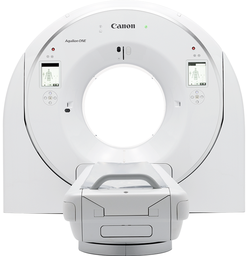
2024 flagship with PUREINSIGHT 320-row detector, 0.241s rotation, and 4D whole-brain perfusion capability.
View Details →Also Serviced & Supported
We service and source parts for GE Healthcare, Siemens, Philips, and other CT platforms. Contact us →
Canon Medical · CT Scanners
Simply delivers.
An 80-row CT scanner designed for departmental efficiency and outstanding image quality. The Aquilion Serve brings AI-powered automation and deep learning reconstruction to workhorse CT operations.
Specifications
| Detector | PUREVision, 80 rows × 0.5 mm |
| Rotation Speed | 0.5 sec standard / 0.35 sec optional |
| Gantry Bore | 80 cm |
| Generator | 50.4 kW |
| kV Selection | 80, 100, 120, 135 kV |
| Table Capacity | 315 kg (694 lbs) |
| Table Height (min) | 33 cm |
| Lateral Slide | 17 cm (8.5 cm each direction) |
| Reconstruction Speed | Up to 70 fps (optional) |
| Installation Space | 14.8 m² minimum |
Key Features
40% reduction in workflow steps. Anatomic landmark detection with 97% accuracy for scan range planning.
Whole-body DLR delivers 1.5 mm low-contrast resolution at 0.3% — clinical-grade image quality at lower dose.
Ultra-low dose lung screening as low as 0.3 mSv. Advanced spectral pre-filtration for dose optimization.
Powered by Canon Medical’s Altivity AI platform for intelligent scan planning, quality assurance, and clinical decision support.
Wide gantry bore with 33 cm minimum table height accommodates a broad range of patient sizes and clinical scenarios.
14.8 m² minimum installation space enables deployment in facilities where larger suites are not feasible.
Contact Rose Rock Biomedical for sales, service, and parts availability.
Canon Medical · CT Scanners
Simply performs.
The premium 80-row performer. With a 72 kW generator, 0.35-second rotation, and the full INSTINX AI suite, the Serve SP delivers exceptional throughput and image quality in a compact, efficient package.
Specifications
| Detector | PUREVision, 80 rows × 0.5 mm |
| Rotation Speed | 0.35 sec |
| Gantry Bore | 80 cm |
| Generator | 72 kW |
| kV Selection | 80, 100, 120, 135 kV |
| Table Capacity | 315 kg (694 lbs) |
| Table Height (min) | 33 cm |
| Lateral Slide | 17 cm (8.5 cm each direction) |
| Reconstruction Speed | Up to 70 fps |
| AiCE Low-Contrast Resolution | 1.5 mm at 0.3% |
| Installation Space | 14.8 m² minimum |
Key Features
High-power generator enables demanding clinical protocols including cardiac and bariatric imaging with consistent output.
Fast gantry rotation reduces motion artifacts and expands cardiac and pediatric imaging capabilities.
40% reduction in workflow steps. 97% accuracy in anatomical landmark detection for automated scan range planning.
Deep learning reconstruction across all body regions — sharp detail, low noise, and reduced dose in one workflow.
Advanced spectral pre-filtration for ultra-low dose lung screening and routine protocols.
Canon’s AI platform powers intelligent workflow, quality monitoring, and clinical decision support throughout the exam.
Contact Rose Rock Biomedical for sales, service, and parts availability.
Canon Medical · CT Scanners
Innovate. Illuminate. Initiate.
A 320-row volume CT scanner delivering 16 cm of simultaneous whole-organ coverage in a single rotation. Spectral CT capability, PIQE 1024 deep learning reconstruction, and dynamic 4D imaging — setting the standard in advanced CT.
Specifications
| Detector | PUREViSION, 320 rows × 0.5 mm |
| Z-Axis Coverage | 16 cm (whole-organ in one rotation) |
| Slices | Up to 640 (with coneXact reconstruction) |
| Rotation Speed | 0.275 sec |
| Gantry Bore | 78 cm |
| Gantry Tilt | ±30° |
| Generator | 100 kW (optional) |
| Table Capacity | 315 kg (694 lbs) |
| Reconstruction Speed | Up to 80 fps |
| Lung Screening Dose | 0.3 mSv (with SilverBeam) |
| Installation Space | 19 m² |
Key Features
Capture the entire heart, brain, or organ in a single rotation — eliminating step-and-shoot artifacts in dynamic studies.
Super-resolution reconstruction delivering exceptional detail and noise reduction across all clinical applications.
Single-source spectral CT for material decomposition, virtual monoenergetic imaging, and iodine quantification.
Time-resolved 4D imaging of the heart, brain perfusion, and joints — all within the 16 cm single-rotation coverage.
AI-guided scan planning, automated positioning, and workflow optimization reduce steps and variability across the team.
Supports real-time CT guidance for biopsy and interventional procedures with integrated 4D visualization.
Contact Rose Rock Biomedical for sales, service, and parts availability.
Canon Medical · CT Scanners
Advanced imaging simplified.
Canon Medical’s 2024 flagship CT — winner of the 2024 Minnies Award for Best New Radiology Device. The PUREINSIGHT 320-row detector with 0.241-second rotation and CLEAR Motion cardiac correction sets a new benchmark for clinical CT.
Specifications
| Detector | PUREINSIGHT, 320 rows × 0.5 mm isotropic |
| Z-Axis Coverage | 16 cm |
| Slices | Up to 640 (coneXact double-slice) |
| Rotation Speed | 0.241 sec |
| Gantry Bore | 80 cm (flared design) |
| Gantry Tilt | ±30° |
| Generator | 100 kW |
| Max Tube Current | 1400 mA |
| kV Selection | 70, 80, 100, 120, 135 kV |
| Table Capacity | 315 kg (695 lbs) |
| Table Height (min) | 33 cm |
| Lung Screening Dose | 0.3 mSv (SilverBeam filter) |
| Installation Space | 19.38 m² minimum |
Key Features
Next-generation detector with isotropic 0.5 mm resolution across 16 cm of coverage — a major leap over the PRISM generation.
Fastest rotation in the Aquilion ONE family — enabling motion-free cardiac imaging without additional heart rate medication.
AI-powered cardiac motion correction reconstructs sharp coronary images even at elevated or irregular heart rates.
40% reduction in electronic noise vs. predecessor, delivering extraordinary spatial resolution via deep learning reconstruction.
16 cm coverage captures complete cerebral perfusion in a single rotation — no toggling, no coverage gaps.
Next-generation tube technology with enhanced heat dissipation for sustained high-output scanning in demanding clinical settings.
Contact Rose Rock Biomedical for sales, service, and parts availability.
X-Ray Systems
Room-based, fluoroscopy, and flat panel DR systems. Select a model below.
Ceiling-mounted manual positioning system with 800 lb table capacity and color-coded rail controls.
View Details →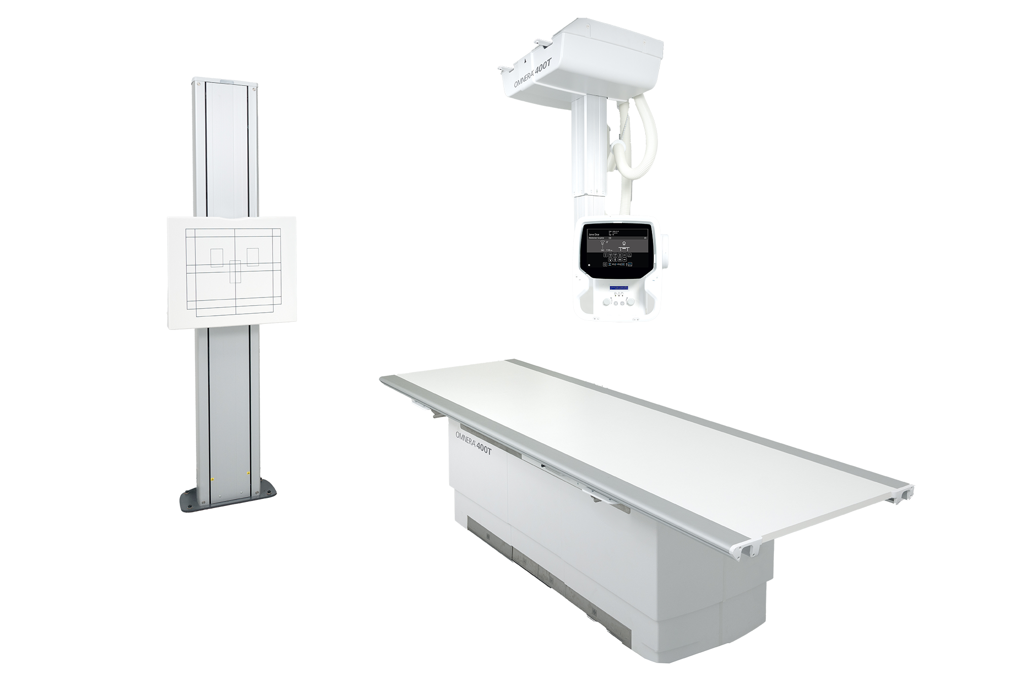
Ceiling-mounted system with selectable detector tracking, 50 kW generator, and 12.1” touchscreen.
View Details →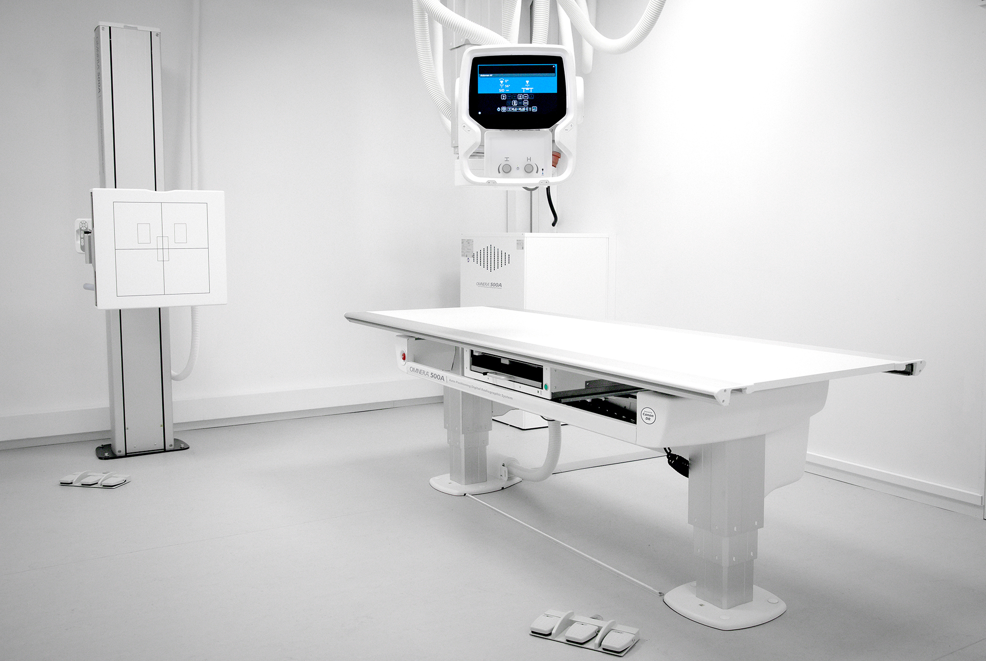
Advanced auto-positioning ceiling-mounted DR with motorized wall stand and enterprise detector sharing.
View Details →
Freestanding floor-mounted system with 9-foot rails and 4-way float table. No structural modifications required.
View Details →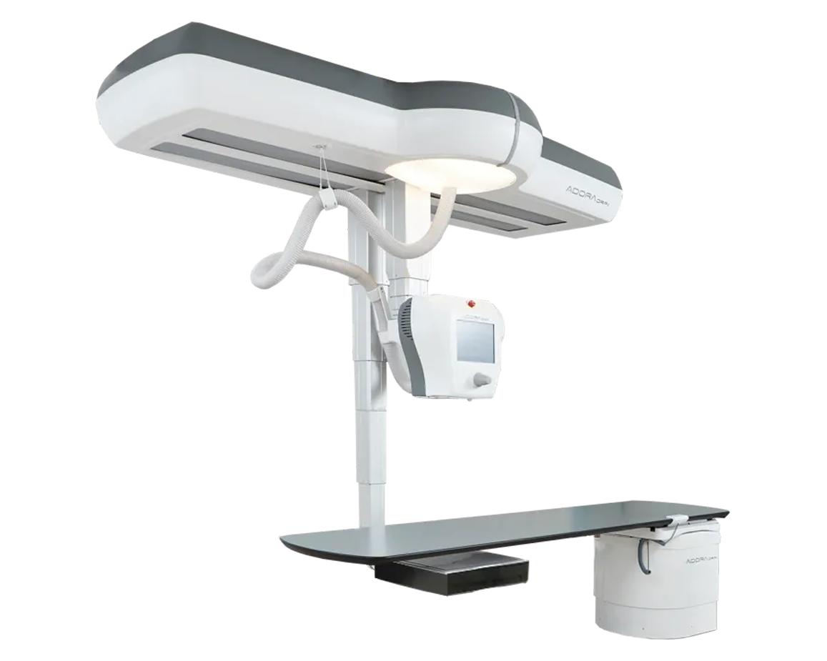
Hybrid radiography and fluoroscopy room with 80 kW generator and ceiling-suspended C-arm. FDA cleared 2024.
View Details →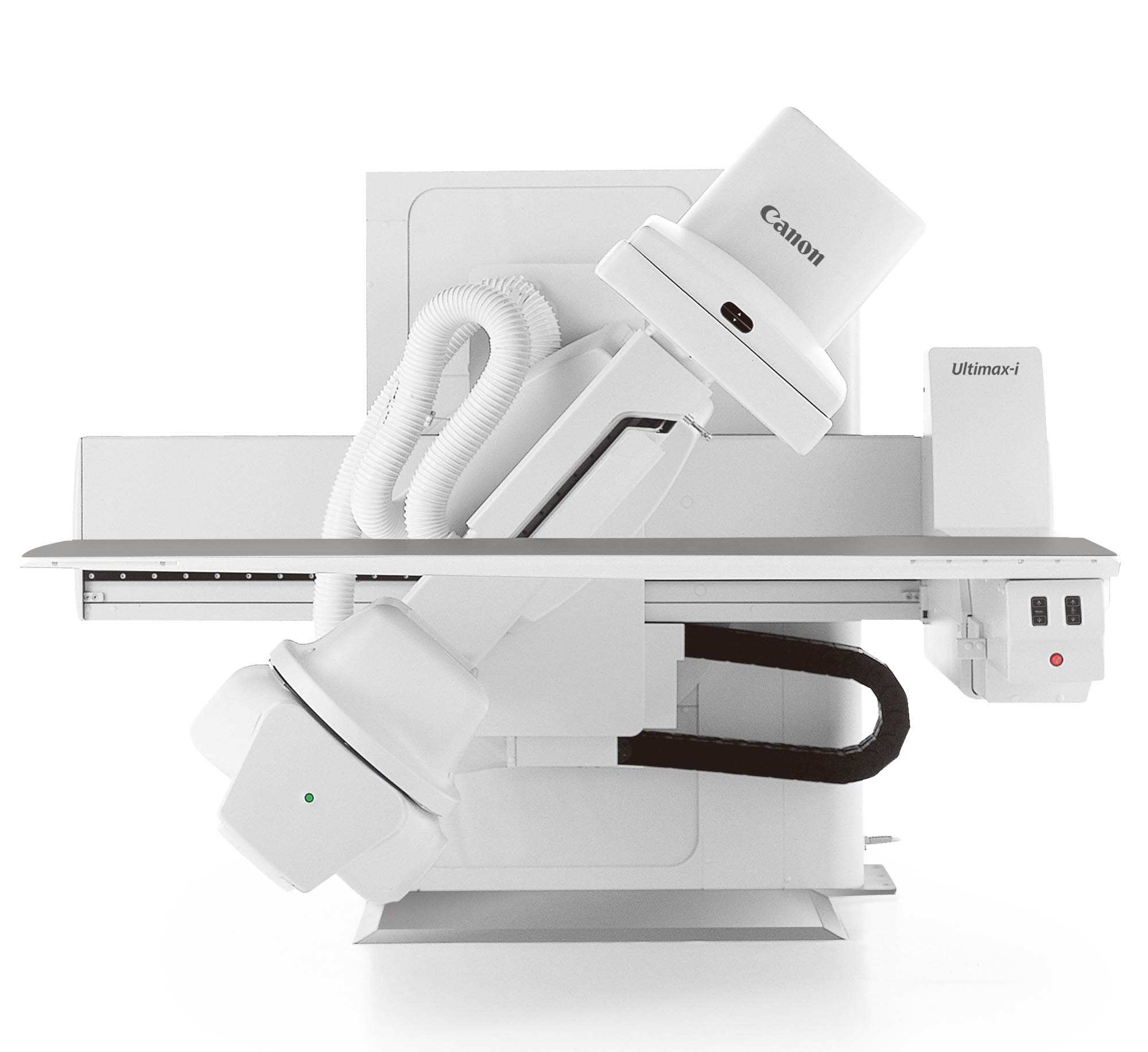
Multipurpose R/F system with ±89° table tilt, multidirectional C-arm, and DoseRite dose management suite.
View Details →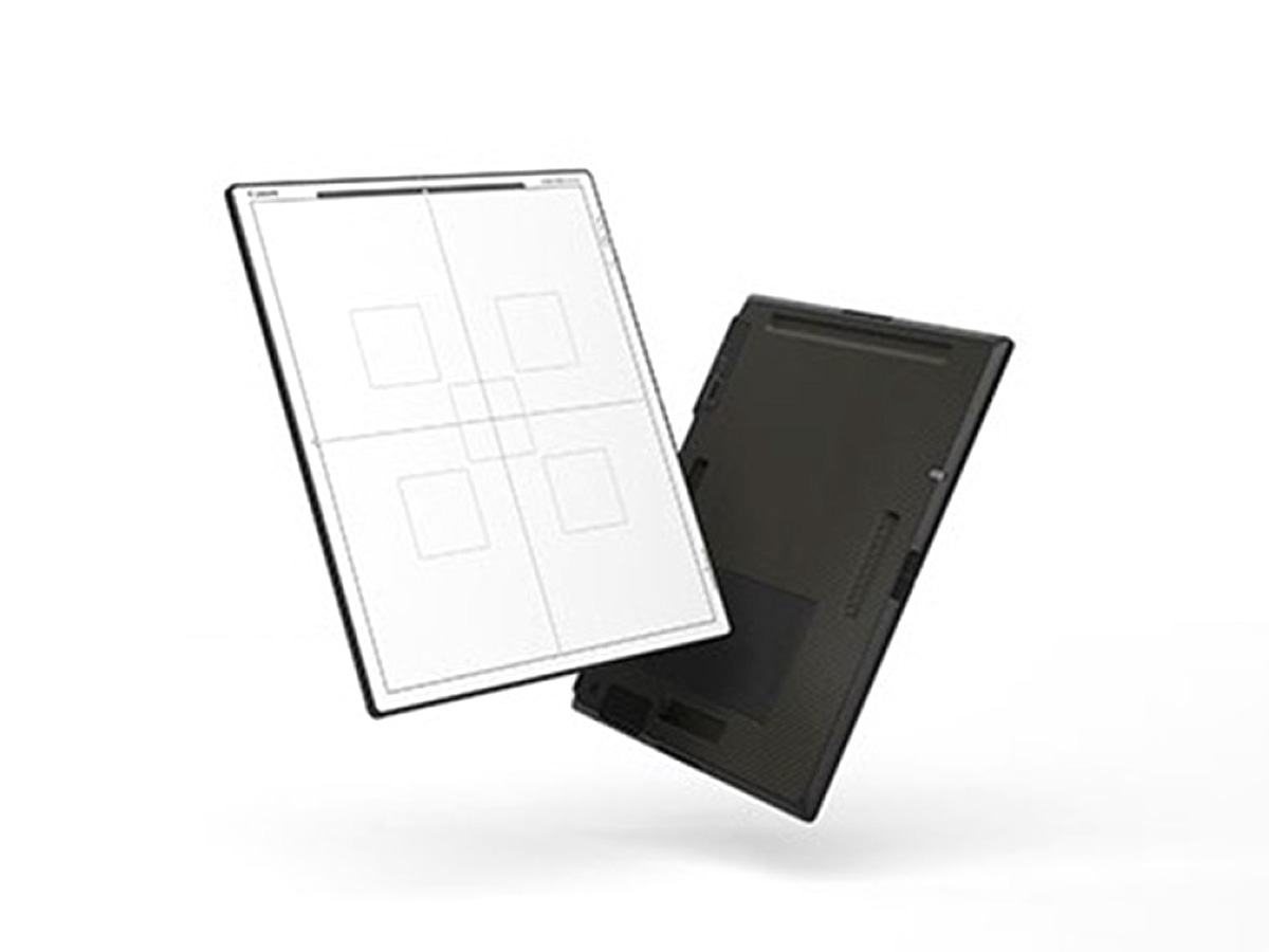
Elite wireless flat panel DR detector. IP57 waterproof, 3-second ready time, 16% DQE improvement.
View Details →Also Serviced & Supported
We service and source parts for GE, Siemens, Philips, Fujifilm, and Carestream X-ray systems. Contact us →
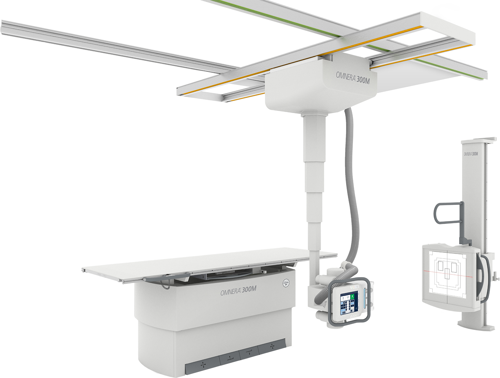
Canon Medical · X-Ray Systems
Ceiling-mounted manual positioning system.
A robust ceiling-mounted radiography system built for high-volume departmental workflows. The OMNERA 300M combines a 5-tier telescoping column, 800 lb table capacity, and intuitive color-coded controls with Canon CXDI digital detector compatibility.
Specifications
| System Type | Ceiling-mounted, manual positioning |
| Table Width | 36 inches |
| Table Capacity | 800 lbs |
| Table Height (min) | 22.25 inches |
| OTC Telescoping Column | 5-tier, 70.8” vertical range |
| Touchscreen | 10.4” (OTC console) |
| Collimator | Manual (auto optional) |
| Brake System | Electromagnetic |
| Detector Compatibility | Canon CXDI digital detectors |
Key Features
Intuitive color-coded ceiling rail controls reduce operator error and speed positioning during busy imaging sessions.
Extra-heavy-duty table accommodates a wide range of patient sizes without requiring a separate bariatric system.
70.8” vertical travel range with electromagnetic braking for precise, repeatable tube positioning at any height.
Completely smooth, flat tabletop surface simplifies cleaning, reduces contamination risk, and improves patient comfort.
Side and overhead patient handgrips plus adjustable lateral support bar for positioning assistance during exams.
Works with the full range of Canon CXDI wireless digital detectors for immediate digital imaging at the table or wall stand.
Contact Rose Rock Biomedical for pricing and availability.
Canon Medical · X-Ray Systems
Selectable tracking system.
A ceiling-mounted DR system with selectable detector tracking to both the table and wall stand. The 50 kW high-frequency generator, 12.1” touchscreen, and optional self-supporting installation cube make it flexible for any department configuration.
Specifications
| System Type | Ceiling-mounted with selectable tracking |
| Generator | 50 kW high-frequency (100–240 kHz) |
| Max kV | 150 kV |
| X-Ray Tube | 400 kHu high-capacity |
| Table Capacity (standard) | 650 lbs |
| Table Capacity (2-column) | 660 lbs |
| Table Type | Flexible elevating 4-way float |
| Touchscreen | 12.1” |
| Detector Compatibility | Canon CXDI digital detectors |
Key Features
Automatic detector tracking to the wall stand or table position streamlines positioning and reduces exam time.
Consistent, precise exposures at 100–240 kHz for demanding exam types with reproducible dose output.
Self-supporting installation cube eliminates the need for ceiling structural work — ideal for retrofit installations.
Integrated detector charging in both the wall stand and table ensures wireless detectors are always ready.
Flexible elevating table with 4-way float top for easy patient positioning and lateral centering during exams.
Available in two height versions with customizable rail lengths to match the specific dimensions of your imaging suite.
Contact Rose Rock Biomedical for pricing and availability.
Canon Medical · X-Ray Systems
Advanced auto-positioning.
Canon Medical’s premium ceiling-mounted DR system with full auto-positioning capability. The 500A pairs a motorized, height-adjustable wall stand, enterprise-grade detector sharing, and LED ambient backlighting for an efficient, modern imaging suite.
Specifications
| System Type | Auto-positioning, ceiling-mounted DR |
| Touchscreen | 12” |
| Wall Stand | Motorized, height-adjustable with tilt |
| Detector Sharing | Enterprise-compatible |
| Stitching | Optional automatic stitching package |
| Software | CXDI Control Software NE |
| Ambient Lighting | LED ambient backlighting |
| Detector Compatibility | Canon CXDI digital detectors |
Key Features
Motorized, programmable tube and detector positioning reduces manual adjustments and standardizes exam setup across operators.
Height-adjustable motorized wall stand with tilt for erect, lateral, and angled projections without patient repositioning.
Share Canon CXDI detectors across multiple rooms and systems within the department for maximum utilization.
Optional package automates full-spine and full-leg stitching from multiple exposures into a single composite image.
Single-click image processing, DICOM integration, and PACS send from a consistent, familiar Canon interface.
Configurable room lighting integrated with the system — enhances patient comfort and room environment during exams.
Contact Rose Rock Biomedical for pricing and availability.
Canon Medical · X-Ray Systems
Floor-mounted radiography system.
A freestanding floor-mounted DR system with 9-foot rails and a 4-way float table — designed for fast installation without ceiling or wall structural modifications. Ideal for facilities needing full radiographic capability without major construction.
Specifications
| System Type | Floor-mounted, freestanding |
| Rails | 9-foot rails |
| Table | 4-way float table |
| Chest Stand | Included |
| Tube Arm | Telescoping, rotating |
| Collimator | Manual with laser light and digital SID/angle display |
| Detector Compatibility | Canon CXDI digital detectors |
| Installation | No ceiling or wall structural work required |
Key Features
Self-supporting floor-mounted structure requires no ceiling rails, wall anchors, or structural modifications — dramatically simplifying installation.
Extended rail length provides ample longitudinal coverage for full-range tube positioning across all standard exam types.
Full float-top patient table enables easy lateral centering and longitudinal adjustment without lifting or repositioning patients.
Upright chest stand is included as standard — supporting erect PA and lateral chest exams in the same room setup.
Tube head controls, tracking, and detector charging are all integrated with auto-collimation for streamlined workflow.
Compatible with the full Canon CXDI wireless detector family for immediate digital imaging and PACS integration.
Contact Rose Rock Biomedical for pricing and availability.
Canon Medical · X-Ray Systems
Hybrid DR & fluoroscopy in one room.
FDA cleared December 2024. The Adora DRFi combines full radiography and fluoroscopy capabilities in a single ceiling-suspended system — eliminating the need for separate DR and fluoro suites. Launched to the US market in March 2025.
Specifications
| Generator | 80 kW (65 kW optional), 100 kHz high-frequency |
| kV Range (Rad) | 40–150 kV |
| kV Range (Fluoro) | 40–125 kV |
| mA Range (80 kW Rad) | 10–1000 mA |
| mA Range (Pulsed Fluoro) | 10–80 mA |
| Primary Detector | CXDI-B1 Dynamic, 42 × 43 cm |
| Table Capacity | 250 kg (551 lbs) |
| Table Material | Carbon fiber |
| Tube/Detector Rotation | ±270° |
| Base Rotation | 340° |
| Auto-Position Memory | Up to 999 positions |
| Touchscreen | 12” floating |
| FDA Status | 510(k) cleared December 2024 |
Key Features
Single detector handles both true dynamic fluoroscopy and static radiography — no detector swap needed during hybrid studies.
Patented ceiling-suspended tube and detector unit enables full rotation and horizontal projections without patient repositioning.
Store and recall up to 999 programmed positions for fast, repeatable setup across diverse exam protocols.
Unique geometry enables axial hip projections without requiring patient repositioning — a significant workflow advantage.
Optional CXDI-RF Wireless B1 Detector for docked or fully wireless use during radiographic portions of the exam.
Lightweight, radiolucent carbon fiber table provides excellent image quality in all projections with minimal artifact.
Contact Rose Rock Biomedical for pricing and availability.
Canon Medical · X-Ray Systems
Multipurpose R/F system.
A full-function radiography/fluoroscopy (R/F) system with a multidirectional C-arm, ±89° table tilt, and a 17″ × 17″ flat panel detector. The Ultimax-i supports everything from routine fluoro to DSA and CT-guided interventions — all from a single, shared generator.
Specifications
| Detector | 17″ × 17″ (43 × 43 cm) FPD, 148 μm pixel pitch, 16-bit |
| Table Capacity (horiz.) | 500 lbs |
| Table Capacity (tilted) | 352 lbs |
| Table Tilt | ±89° |
| Table Width | ~20.5 in (60 cm) |
| Table Height (min) | ~20.5 in (52 cm) |
| Longitudinal Range | ~81 in (206 cm) |
| Transverse Movement | ~18 in (48 cm) |
| C-Arm Oblique Rotation | 131° |
| CRA/CRU Angles | 45° |
| SID Range | 34.6″ to 48.4″ |
| X-Ray Tube | Liquid Metal Bearing, 300 KHU |
Key Features
Single generator drives both X-ray tubes and both detectors — simplifying service and reducing total cost of ownership.
Nine integrated dose management technologies including tantalum filtering and virtual collimation minimize patient and staff dose.
In-tube grid switching eliminates soft X-ray leading and trailing edges — improving image quality in pulsed fluoroscopy mode.
Super Noise Reduction Filter provides real-time per-frame noise reduction during fluoroscopy for clean, diagnostic-quality images.
Digital subtraction angiography mode for vascular studies and interventional procedures within the same R/F suite.
Optional WorkRite package for ergonomic C-arm positioning — reducing operator fatigue during long fluoroscopy procedures.
Contact Rose Rock Biomedical for pricing and availability.
Canon Medical · X-Ray Systems
Elite wireless flat panel DR detector.
A next-generation flat panel digital radiography detector with 16% DQE and 29% MTF improvements over previous models. IP57 waterproof, ultra-lightweight, and compatible with any Canon imaging system — the ideal upgrade for any DR workflow.
Specifications
| Model CXDI-720CW | 14″ × 17″ | 2800 × 3408 matrix | 5.1 lbs |
| Model CXDI-820CW | 11″ × 14″ | 3408 × 3408 matrix | 4.0 lbs |
| Model CXDI-420CW | 17″ × 17″ | 2192 × 2800 matrix | 6.0 lbs |
| Pixel Size (all models) | 125 μm |
| Scintillator | CsI (cesium iodide) |
| DQE at 0 lp/mm | 74% (16% improvement over prior gen) |
| DQE at 0.5 lp/mm | 67% |
| Resolution | 4.0 lp/mm |
| IP Rating | IP57 (waterproof) |
| Ready Time | 3 seconds |
| Battery Life | ~2000 images per charge |
| Charge Time | ~150 minutes |
Key Features
Significant detective quantum efficiency gain vs. prior-generation CXDI detectors — more signal per dose at the detector.
Sharper resolving power at 2 lp/mm delivers more detail in fine structures like trabecular bone and small lesions.
Protected against dust and submersion — enabling thorough disinfection and worry-free use in any clinical environment.
Instant-on from standby eliminates wait time during busy workflows — ready as soon as the patient is positioned.
High-capacity battery supports a full clinical day without mid-day charging interruptions.
14×17, 11×14, and 17×17 formats available to match your clinical requirements and patient population.
Contact Rose Rock Biomedical for pricing and availability.
Mobile Radiography
Mobile radiographic systems for bedside imaging in hospitals, clinics, and long-term care facilities.
Canon Medical
Imaging mobility made possible. Pressure-sensitive steering and integrated CsI wireless detector.
View Details →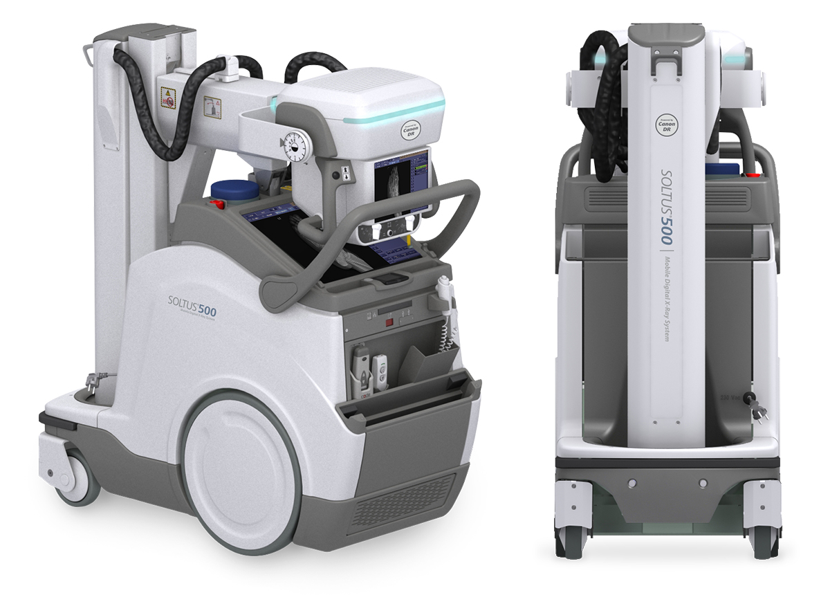
Canon Medical
Bold performance. Compact footprint. 40 kW generator with telescopic column and smart ID card login.
View Details →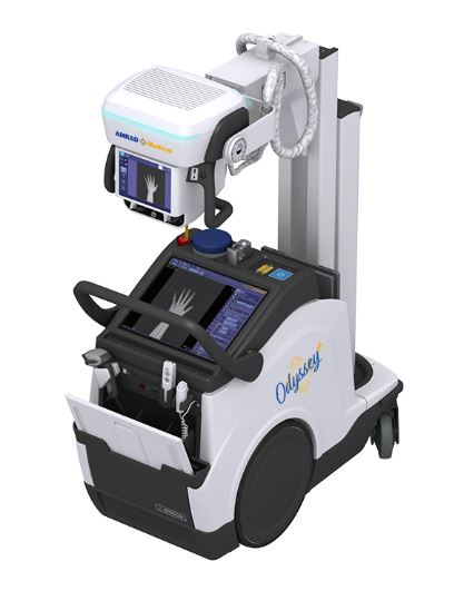
AMRAD Medical
High-frequency mobile X-ray generator with battery operation for complete portability in any care setting.
View Details →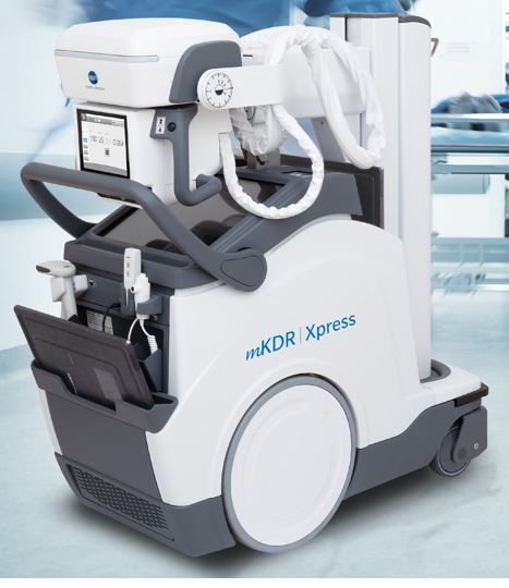
Konica Minolta
Mobile digital radiography system with wireless flat panel detector and intuitive bedside workflow.
View Details →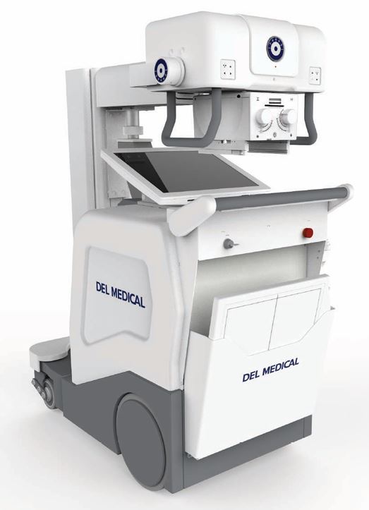
UMG / DEL Medical
Compact, full-featured mobile DR system built for reliable performance across all patient care environments.
View Details →Canon Medical · Mobile X-Ray
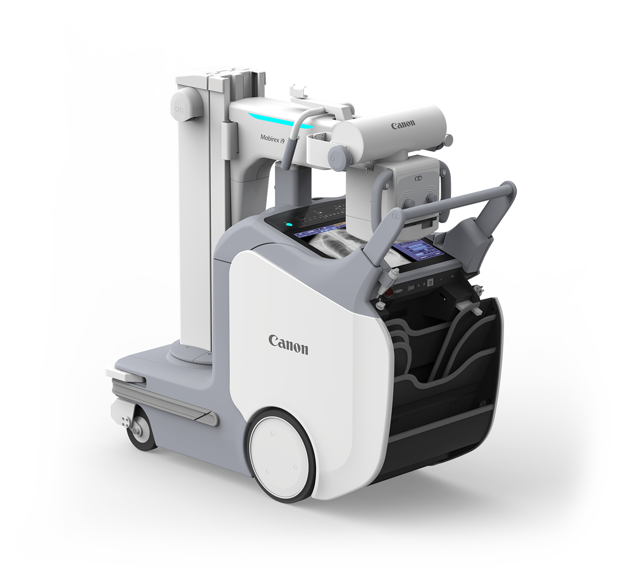
Imaging mobility made possible.
A mobile X-ray system engineered for effortless hospital navigation. Pressure-sensitive steering, an ultra-lightweight integrated CsI wireless detector, and 2-second image display make the Mobirex i9 a standout performer for daily mobile rounds.
Specifications
| Detector Type | CsI flat panel, wireless, waterproof, integrated battery |
| Pixel Size | 125 μm |
| AEC Regions | 5 or 9 (model-dependent) |
| Image Display Time | 2 seconds after exposure |
| Load Capacity | 310 kg (uniform) |
| Unit Width | 560 mm |
| Telescopic Arm Reach | 1200 mm |
| Drive Controls | Pressure-sensitive steering |
| Software | CXDI Control Software NE |
Key Features
Intuitive drive system responds to natural hand pressure — reducing operator fatigue and improving maneuverability in tight spaces.
Ultra-lightweight CsI flat panel with built-in battery is ready at the bedside with no cable management required.
Images appear on the console within 2 seconds of exposure — supporting rapid image review and repeat exposures if needed.
1200 mm reach with telescoping column provides full positioning flexibility for chest, abdomen, and cross-table lateral exams.
Built-in AEC (automatic exposure control) assistance reduces technique variability and improves consistency across operators.
CXDI Control Software NE provides one-click image processing, DICOM send, and dose structured reporting from the mobile console.
Contact Rose Rock Biomedical for pricing and availability.
Canon Medical · Mobile X-Ray
Bold performance. Compact footprint.
A compact 40 kW mobile X-ray system with a telescopic column, smart ID card login, and distributed antenna system for reliable wireless connectivity. Designed for facilities that need dependable mobile DR in a smaller, lighter package.
Specifications
| Generator | 40 kW standard |
| Column | Telescopic |
| Detector | Canon CXDI wireless flat panel |
| Wireless System | Distributed Antenna System (exclusive) |
| Login | Smart ID card system |
| Drive Controls | Secondary controls on collimator handles |
| Software | CXDI Control Software NE |
| Features | Single-click processing, free rotation, scatter correction, stitching |
Key Features
Adequate power output for all standard mobile projections — chest, abdomen, pelvis, and extremity exams across patient populations.
Exclusive wireless antenna design maintains reliable detector connectivity throughout multi-floor hospital environments.
Technologist ID card authentication streamlines access control, enables per-operator exposure tracking, and simplifies compliance.
Secondary drive controls on the collimator handles allow fine positioning adjustments without stepping away from the patient.
Software-based scatter correction improves contrast and detail in chest and abdomen images without a physical grid.
Optional stitching package combines multiple exposures into long-bone or spine composites directly on the mobile console.
Contact Rose Rock Biomedical for pricing and availability.
AMRAD Medical · Mobile X-Ray
Mobile Radiography. No Compromise.
A high-frequency mobile X-ray generator designed for demanding hospital environments. Battery-powered operation enables true cordless mobility from ICU to emergency to long-term care.
Key Capabilities
Fully self-contained battery system enables cordless operation throughout a facility — no searching for outlets in critical care areas. Onboard charging keeps the unit ready between shifts.
High-frequency X-ray generation delivers consistent, reproducible exposures with reduced patient dose — critical for daily mobile rounds across ICU, ED, and post-surgical patients.
Works with a wide range of digital radiography flat panel detectors — wired and wireless — giving facilities flexibility to pair with existing DR infrastructure or upgrade as needed.
Contact Rose Rock Biomedical for pricing, demos, and availability.
Konica Minolta · Mobile X-Ray
Mobile Digital Radiography. Faster Workflow.
An integrated mobile DR system combining a high-output generator with a wireless flat panel detector — delivering immediate diagnostic-quality images at the point of care with minimal setup time.
Key Capabilities
High-resolution amorphous silicon flat panel detector with wireless data transmission — no cables at the bedside. Images available on the console display within seconds of exposure.
Powerful mobile generator ensures consistent exposure quality across all projections — from thin pediatric to large adult patients — without technique compromise.
Intuitive touchscreen interface, anatomical technique programming, and immediate image review keep bedside exams moving without delays or repeat exposures.
Contact Rose Rock Biomedical for pricing, demos, and availability.
UMG / DEL Medical · Mobile X-Ray
Reliable Mobile DR for Any Care Setting.
A full-featured mobile digital radiography system engineered for dependable performance in hospitals, skilled nursing facilities, and outpatient clinics — with the versatility to handle the full range of bedside exam types.
Key Capabilities
Compatible with wired and wireless flat panel detectors for the full spectrum of mobile projections — chest, abdomen, pelvis, extremities, and lateral decubitus — across all patient populations.
Consistent, precise exposures from a proven high-frequency generator platform — delivering reliable image quality with repeatable dose output for daily mobile rounds.
Wi-Fi and Ethernet connectivity with DICOM worklist, PACS send, and dose structured reporting — keeping mobile images in the imaging workflow without extra steps.
Contact Rose Rock Biomedical for pricing, demos, and availability.
Ultrasound Systems
Sales, service & parts for Canon Medical Aplio systems. Select a model below.
Premium general imaging with iBeam architecture, SMI microvascular technology, and Smart Fusion navigation.
View Details →Ideal for routine imaging with full cardiology support, aBeam architecture, and comprehensive elastography suite.
View Details →Mobile, lightweight, multi-purpose — with integrated battery option and smart workflow automation.
View Details →Also Serviced & Supported
We service and source parts for GE Healthcare, Philips, Siemens, Mindray, Sonosite and other ultrasound platforms. Contact us for details →
Canon Medical · Ultrasound
Intuitive. Intelligent. Innovative.
Designed to deliver outstanding clinical precision and departmental productivity. Crystal-clear images with enhanced resolution and penetration.
Key Advantages
Aplio iBeam architecture with dramatically increased processing power provides outstanding imaging clarity while significantly enhancing penetration — from the smallest to the toughest patients.
SMI expands the range of visible blood flow to visualize low-velocity microvascular flow never before seen with diagnostic ultrasound. Separates flow from overlaying tissue motion with superb detail.
Merges real-time ultrasound with previously acquired CT, PET-CT, MR or ultrasound data. Navigate complex anatomy securely and compare lesions with ease across multiple imaging modes.
Technologies
Outstandingly smooth images with sharpened outline of anatomical structures and lesions — even in high BMI patients.
Compounding technology delivering increased imaging contrast and reduced speckle noise to improve visualization.
Increases clinical accuracy and reveals more detail in all depths by electronically sharpening the imaging slice thickness.
Provides harmonic images of extraordinary spatial resolution alongside greatly enhanced penetration.
Comprehensive contrast-enhanced ultrasound imaging and quantification package for perfusion dynamics assessment.
Quantitative measure and real-time display of tissue elasticity with unique propagation map for quality assessment.
Clear visualization of biopsy needles in the live image. Works with all common needle sizes and selects optimal enhancement automatically.
Simple volume-imaging capability for convex and linear transducers, supporting all modes including SMI and shear wave.
Ultra-Wideband Transducers
Contact Rose Rock Biomedical for sales, service, and parts availability.
Canon Medical · Ultrasound
Advanced. Integrated. Seamless.
Designed to enhance productivity and throughput while maximizing clinical confidence. The ideal system for routine imaging departments with an extensive selection of advanced applications.
Key Advantages
Unique aBeam architecture ensures all imaging technologies work together seamlessly for greater uniformity across all applications — reducing clutter, strengthening signal and improving visualization.
Wall Motion Tracking provides immediate visual and quantitative access to global and regional myocardial dynamics. Comprehensive stress echo support for physical and pharmacological protocols.
Comprehensive shear wave and strain elastography suite with raw data functionality. Smart Maps visualize and quantify shear wave propagation in real time.
Technologies
Outstandingly smooth images with sharpened outline of lesions, enhanced uniformity and reduced clutter.
Compounding technology delivering increased imaging contrast and reduced speckle noise.
Color-coded SMI depicts flow and greyscale information with high temporal and spatial resolution simultaneously.
High spatial resolution color Doppler to reveal minute flow patterns with extraordinary accuracy and detail.
Comprehensive contrast-enhanced ultrasound package for perfusion dynamics across a wide range of clinical settings.
Merges real-time ultrasound with previously acquired CT, MR or ultrasound data in a simple two-step process.
Natural-looking 3D renderings with strong visual feedback on depth and detail from first trimester.
High frame rate Tissue Doppler images and PW-TDI traces for precise timing of cardiac events.
Clinical Applications
Contact Rose Rock Biomedical for sales, service, and parts availability.
Canon Medical · Ultrasound
Scan smart. Ignite your potential.
Pristine images, automated functions, intelligent workflows. The ideal system to handle a wide variety of imaging needs in a busy clinic — with outstanding mobility and versatility.
Key Advantages
Small form factor, low weight, and large easy-roll casters make the system extremely maneuverable. The fully integrated battery option supports remote, cordless operation for up to 30 minutes.
Consistently high performance across common and specialized clinical applications. Many applications can be fully covered with just one of Canon’s high-performance transducers — reducing cost and simplifying workflow.
Protocol Assistant navigates you through exams with a clear, easy-to-read menu. Connects seamlessly into hospital networks with PACS/HIS/RIS integration.
Technologies & Connectivity
Remarkably smooth images with sharp outlines, improved uniformity and reduced clutter — rock-solid image quality every exam.
Elevates color Doppler to reveal minute blood flow with exceptional detail and sensitivity.
Unique application for visualizing and quantifying the attenuation coefficient — useful for assessing fatty liver disease.
Smooth, natural and detailed 3D/4D rendering with strong visual feedback on depth and detail across all clinical applications.
Seamless PACS/HIS/RIS integration. Optional Tricefy cloud services for instant image sharing. ApliGate for secure remote consultation.
Next-generation ergonomic transducers with outstanding clinical versatility, thin super-flexible cables, and color-coded connector identification.
Clinical Applications
Contact Rose Rock Biomedical for sales, service, and parts availability.
Vascular Diagnostics
Peripheral vascular assessment systems for pressure measurement, blood flow analysis, and tissue oxygenation.
Non-invasive ABI and segmental limb pressure measurement for peripheral arterial disease assessment.
View Details →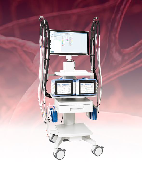
Multi-modality vascular assessment combining pressure and laser Doppler flowmetry in one platform.
View Details →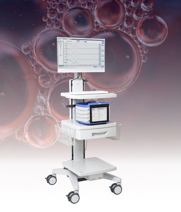
Transcutaneous oxygen monitoring for wound care assessment, limb salvage, and hyperbaric therapy planning.
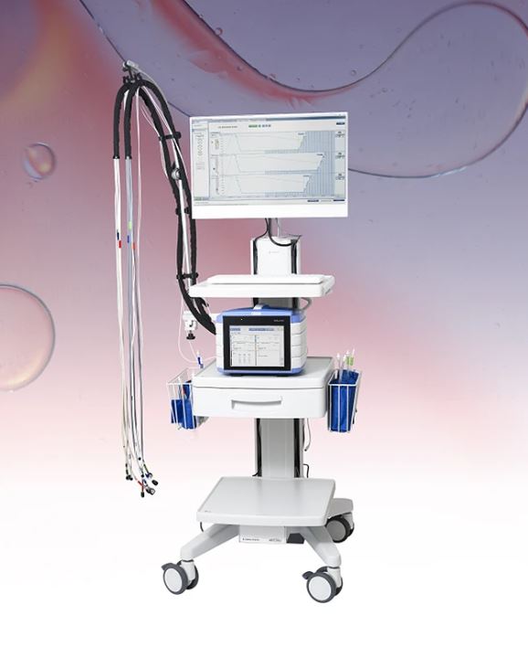
View Details →Perimed · ABI Systems
Non-Invasive Peripheral Pressure Measurement
A complete system for ankle-brachial index (ABI) measurement, segmental limb pressures, and pulse volume recording — the essential tools for diagnosing and monitoring peripheral arterial disease.
Key Capabilities
Calculates the ankle-brachial index automatically from simultaneous limb pressure measurements. Standardized protocols for consistent, reproducible results across examiners and visits.
Multi-level cuff measurements at the thigh, calf, and ankle to pinpoint the location of arterial disease and assess disease severity throughout the lower limbs.
Non-invasive plethysmographic waveform analysis provides qualitative blood flow information to complement pressure measurements — essential for borderline ABI results.
System Features
High-sensitivity continuous wave Doppler probe for precise Doppler signal acquisition at the ankle and toe.
Photoplethysmography (PPG) probes for toe systolic pressure — critical for diabetic patients where ABI may be falsely elevated.
Automated multi-channel cuff inflation and deflation reduces examiner variability and improves patient comfort during testing.
Post-exercise ABI testing on a treadmill to detect exercise-induced ischemia in patients with normal resting ABI.
Real-time waveform display with automatic classification of waveform morphology — triphasic, biphasic, or monophasic.
Professional PDF report generation with waveforms, pressure indices, and interpretation guidance ready for physician review.
Clinical Applications
Contact Rose Rock Biomedical for pricing and availability.
Perimed · ABI Systems
Multi-Modality Vascular Assessment
Combines peripheral pressure measurement with laser Doppler flowmetry in a single integrated platform — giving vascular labs the breadth of assessment they need without multiple devices.
Key Capabilities
Simultaneously captures peripheral blood pressure indices and microvascular laser Doppler flowmetry data — providing a comprehensive picture of both macro and microcirculation in a single exam session.
Real-time, non-invasive measurement of cutaneous blood flow using laser light. Ideal for assessing microvascular function, reactive hyperemia, and endothelial response to vasoactive stimuli.
Comprehensive evaluation of tissue perfusion and viability — combining pressure indices with microvascular flow data to support wound care planning, amputation level determination, and treatment monitoring.
Contact Rose Rock Biomedical for pricing and availability.
Perimed · ABI Systems
Transcutaneous Oxygen Monitoring
Non-invasive measurement of tissue oxygen tension (TcpO2) for wound care assessment, limb salvage planning, and hyperbaric oxygen therapy monitoring — delivering critical perfusion data where it matters most.
Key Capabilities
Heated skin surface electrodes measure the partial pressure of oxygen diffusing through the skin — providing a direct, real-time indicator of tissue perfusion and oxygen delivery at the measurement site.
TcpO2 values predict wound healing potential and guide treatment decisions — identifying patients who require revascularization before wound closure or amputation level determination.
In-chamber TcpO2 monitoring with optional hyperbaric-rated probes confirms adequate oxygen delivery during HBOT sessions and identifies patients likely to respond to hyperbaric oxygen treatment.
System Features
Monitor up to four sites simultaneously — comparing contralateral limbs or tracking multiple wound sites in a single session.
Precisely temperature-controlled Clark-type electrodes ensure consistent, reproducible oxygen measurements at the skin surface.
Optional probes rated for use inside hyperbaric chambers — supporting in-chamber response testing under elevated pressure.
Continuous trend display with configurable alarms for low TcpO2 thresholds — ideal for long-term monitoring in wound care settings.
Automatic calculation of the oxygen tension index (OI) for standardized assessment of regional perfusion against a reference site.
Comprehensive report output with trend graphs, threshold annotations, and clinical decision support data for physician review.
Clinical Applications
Contact Rose Rock Biomedical for pricing and availability.